

Rs 567 only/-
9907385555 asingh039@gmail.com
MP NAGAR BHOPAL
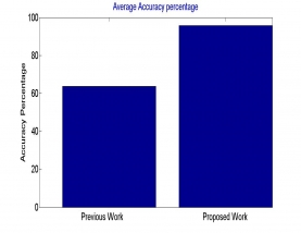
Brain tumor identification is tricky assignment because of complex structure of human brain. X-ray pictures created from MRI scanners utilizing solid Magnetic fields and radio waves to shape pictures of the body which helps for medicinal finding. This paper present the MRI picture of brain tumor into two class initially is tumor region while other is non tumor one. Here by utilizing Active Contour calculation segmentation of tumor area is possible with no earlier preparing with high precision. Proposed calculation use median filter to remove the noise part of the picture. Investigation was done on genuine picture dataset. Results are compared with existing strategies on different assessment parameters and it was discovered that proposed calculation was superior.
Medicinal picture analysis[2] can be utilized as initial screening strategies to help specialists. Different parts of segmentation features and calculations have been broadly investigated for a long time in a large group of productions. In any case, the issue stays hard, with no broad and special arrangement, because of an expansive and continually developing number of various objects of intrigue, huge varieties of their properties in pictures, distinctive medical imaging modalities, and related changes of flag homogeneity, inconstancy, and clamor for each question. Computed Tomography (CT) and Magnetic Resonance(MR) imaging are the most generally utilized radiographic methods in analysis, clinical investigations and treatment arranging.
In this process picture is resize in settle measurement. As various picture have distinctive measurement. So change of each is done in this progression. One more work is to change over all picture in gray level. As various picture has RGB, HSV, and so forth arrange so working at single configuration is required.
| IEEE Base paper | |||
| Doc | Complete Project word file document | ||
| Read me | Complete read me text file | ||
| Source Code | Complete Code files |