


Rs 567 only/-
9907385555 deepaktest.patel@gmail.com
MP NAGAR BHOPAL
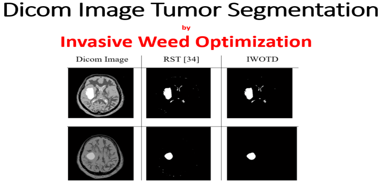
This work takes brain computer aided diagnosis (CAD) images as input and identify the tumor region in the image. Preprocessing steps of tumor detection increase the final accuracy of models. Pre-processing of input image was done by Computer-Aided Diagnosis in which a second-order statistic filter was applied for noise removal and skull pixel extraction. Input image noise and skull play an adverse effect on tumor diagnosis, so this needs to be preprocessed before detection. Dynamic adoption of genetic algorithm does not need any prior training or information. This work has applied Invasive Weed Optimization Tumor Detection algorithm for brain tumor detection from Dicom images. Invasive Weed Optimization algorithm for identifying the cluster center pixels in the image. Finally, the image gets clustered into the tumor and non-tumor regions. The experiment was done on a real dicom image dataset.
| IEEE Base Paper | |||
| Doc | Document File | ||
| Source Code | Complete Code Files |
