

Rs 648 only/-
9907385555 jaipur.researchs@gmail.com
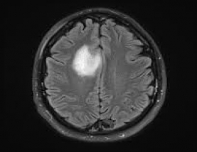
Abstract: Brain tumor identification is tricky assignment because of complex structure of human brain. X-ray pictures created from MRI scanners utilizing solid Magnetic fields and radio waves to shape pictures of the body which helps for medicinal finding. This paper present the MRI picture of brain tumor into two class initially is tumor region while other is non tumor one. Here by utilizing Active Contour calculation segmentation of tumor area is possible with no earlier preparing with high precision. Proposed calculation use median filter to remove the noise part of the picture. Investigation was done on genuine picture dataset. Results are compared with existing strategies on different assessment parameters and it was discovered that proposed calculation was superior.
Methodology: This work focus on the digital MRI image segmentation. Whole work was divide into three step first was pre-processing in this step noise was detect than remove from the input image which was due to the salt or pepper attack. Second step was to segment image by using Active contour method into few region termed as skull, brain and tumor. Third step was to remove skull part from the image and finally highlight tumor portion of the image.
Results: MATLAB 2012a was the software use for the implementation of this work. Results shows that proposed work has achieved a high precision recall, and F-Measure value with as the testing files as compare to previous existing methods. It was achieved because of active contour segmentation.
Keywords: Active Contour, Denoising, Median Filter, MRI image, Segmentation, Unsupervised.
Objective
| Base Paper | |||
| Doc | Complete Document | ||
| Source Code | Complete Code files |